231/3 · BIOLOGY (Practical) · Paper 3 · Dec. 2022 · 1¾ hours
Instructions: Answer all the questions in the spaces provided. Spend the first 15 minutes reading the whole paper carefully before commencing your work.
Question 1
You are provided with: a prepared slide, labelled E, containing a transverse section through a plant organ; access to a light microscope with at least the low and medium power objective lenses.
(a) Observe the section under the low power and medium power objective lenses of the light microscope.
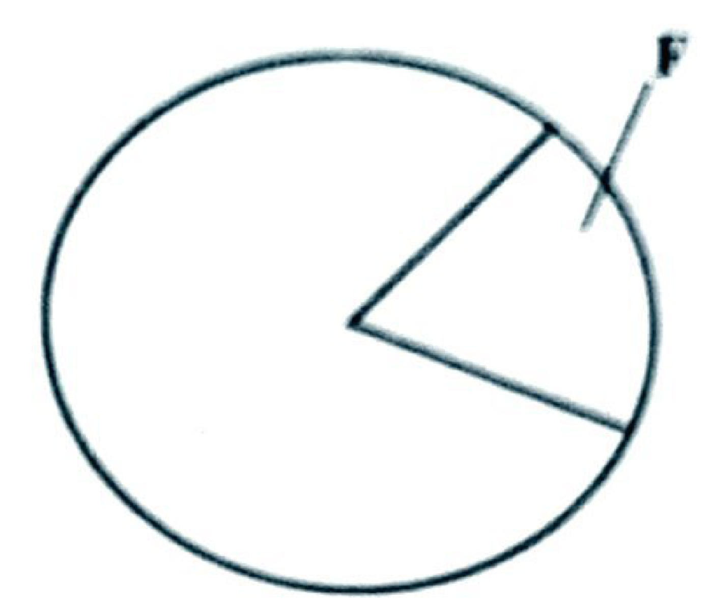
(i) Fill the portion labelled F in the plan diagram below to show (a portion of) the structures seen under the medium power objective of the light microscope. Label the structures. (3 marks)
(ii) Calculate the magnification of the image observed under the low power objective lens. (2 marks)
(b) With reference to one observable feature, state the class of plants from which the organ was obtained. (2 marks)
(c) Name the plant part from which the section was obtained. (1 mark)
(d) State two precautions one should take to ensure the safety of the slide during observation under the microscope. (2 marks)
(e) State how each of the following parts of the light microscope contributed to the clarity of the image of the section observed: (i) mirror; (ii) diaphragm; (iii) condenser. (3 marks)
Question 2
You are provided with: half a portion of raw banana; three beakers labelled G, H and J — G contains 50 ml of dilute hydrochloric acid, H contains 50 ml of distilled water, J is empty; a scalpel; a spatula/pair of forceps; a white plain paper or white tile; a stopwatch/means of timing.
When some plant tissues are exposed, enzymes on the exposed surfaces react with oxygen. Using the provided materials, investigate the enzyme–oxygen reaction using the procedure below.
I. Slice off about 2 mm from the exposed end of the raw banana and discard the slice.
II. Slice another piece, about 1 cm wide, from the remaining banana to use in the investigation.
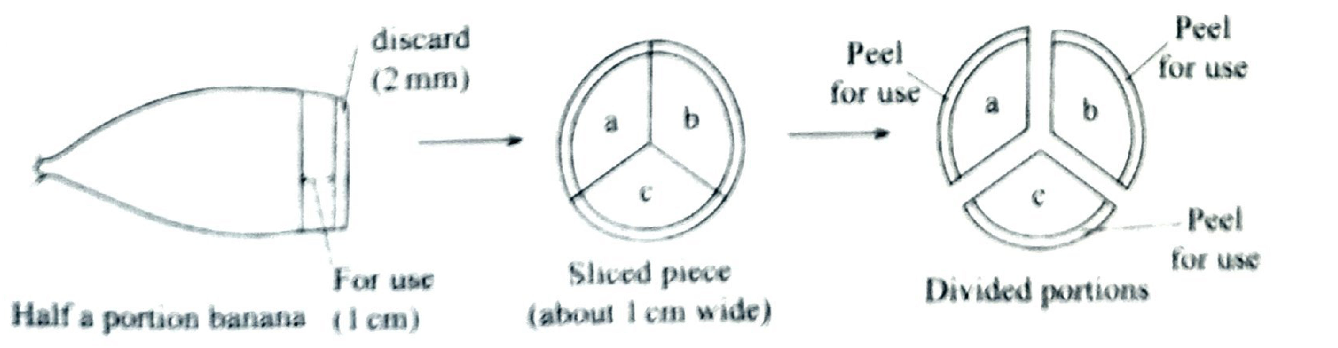
III. Divide the portion obtained in (II) into three parts (a, b and c) as illustrated in the diagram below.
IV. Remove the peel from portion a, cut the peel into three pieces and immediately drop all three pieces into beaker G (containing hydrochloric acid). Obtain peels only, without remnants of banana flesh.
V. Repeat procedure IV with the peels from portion b into beaker H (containing distilled water) and those from portion c into beaker J.
VI. Leave the set-up for five minutes and observe the inner surfaces of the banana peels in each beaker.
(a) (i) Record the observations made in each case: G, H, J. (3 marks)
(ii) Account for the observations made in beakers H (2 marks) and J. (2 marks)
VII. Using the spatula/pair of forceps, remove the peels from each beaker and expose the sets of peels separately on the plain paper/white tile. Leave them for a further five minutes and observe.
(b) (i) Record the observations made on the peels from beakers H (1 mark) and J. (1 mark)
(ii) Account for the observations on the surfaces of the peels from beakers G (1 mark) and J (1 mark) after a further 5 minutes.
(c) Suggest the suitable pH for the enzymes found on the surface of the banana peels. (1 mark)
(d) Suppose the peels in set-up J were initially boiled for 5 minutes. (i) Suggest the observations that would have been made. (1 mark) (ii) Explain the observations made in (d)(i). (1 mark)
Question 3
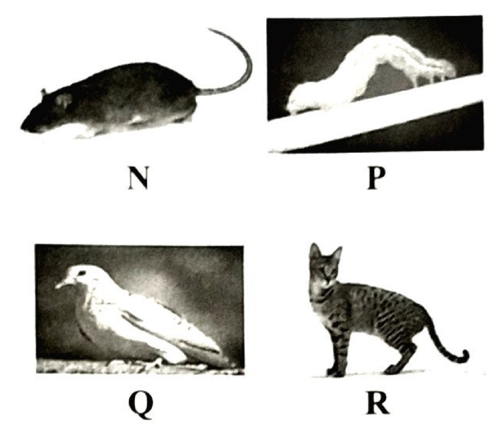
You are provided with specimens labelled K and L together with photographs of organisms labelled N, P, Q and R.
Assuming the organisms are found in the same ecosystem:
(a) (i) Construct a complete food web that includes the specimens and photographs of organisms in this ecosystem. (8 marks)
(ii) From the food web, identify the longest food chain. (2 marks)
(b) (i) Identify the organisms with the highest biomass. (1 mark) (ii) Give a reason for your answer in (b)(i). (1 mark)
(c) Other than feeding, explain how the organism represented in photograph R benefits from specimen K. (1 mark)